Humerus
An anatomical and biomechanical study guide focusing on the long bone of the upper arm — its bony landmarks, muscular attachments, and clinical significance.
1. Osteology of the Humerus
Divided into proximal epiphysis, diaphysis (shaft), and distal epiphysis
| Region | Landmark |
|---|---|
Proximal | Head |
| Anatomical Neck | |
| Surgical Neck | |
| Greater Tubercle | |
| Lesser Tubercle | |
| Intertubercular Groove | |
Shaft | Deltoid Tuberosity |
| Radial (Spiral) Groove | |
Distal | Medial Epicondyle |
| Lateral Epicondyle | |
| Capitulum | |
| Trochlea | |
| Olecranon Fossa | |
| Coronoid Fossa |
2 & 3. Muscular Attachments
Muscles originating from and inserting onto the humerus
These muscles have their proximal attachment on the humerus and typically act on the elbow or forearm.
| Muscle | Origin Site |
|---|---|
| Brachialis | Distal half of anterior humerus |
| Brachioradialis | Lateral supracondylar ridge |
| Triceps Brachii (Medial & Lateral Heads) | Posterior surface of humerus |
| Anconeus | Lateral epicondyle |
| Common Flexor Tendon | Medial epicondyle |
| Common Extensor Tendon | Lateral epicondyle |
| Supinator | Lateral epicondyle (and ulna) |
These muscles originate on the axial skeleton or scapula and act primarily on the glenohumeral joint (shoulder).
| Muscle | Insertion Site |
|---|---|
| Supraspinatus | Greater tubercle (superior facet) |
| Infraspinatus | Greater tubercle (middle facet) |
| Teres Minor | Greater tubercle (inferior facet) |
| Subscapularis | Lesser tubercle |
| Pectoralis Major | Lateral lip of the bicipital groove |
| Latissimus Dorsi | Floor of the bicipital groove |
| Teres Major | Medial lip of the bicipital groove |
| Deltoid | Deltoid tuberosity |
| Coracobrachialis | Medial aspect of the mid-shaft |
4. Functional Mechanics & Force Couples
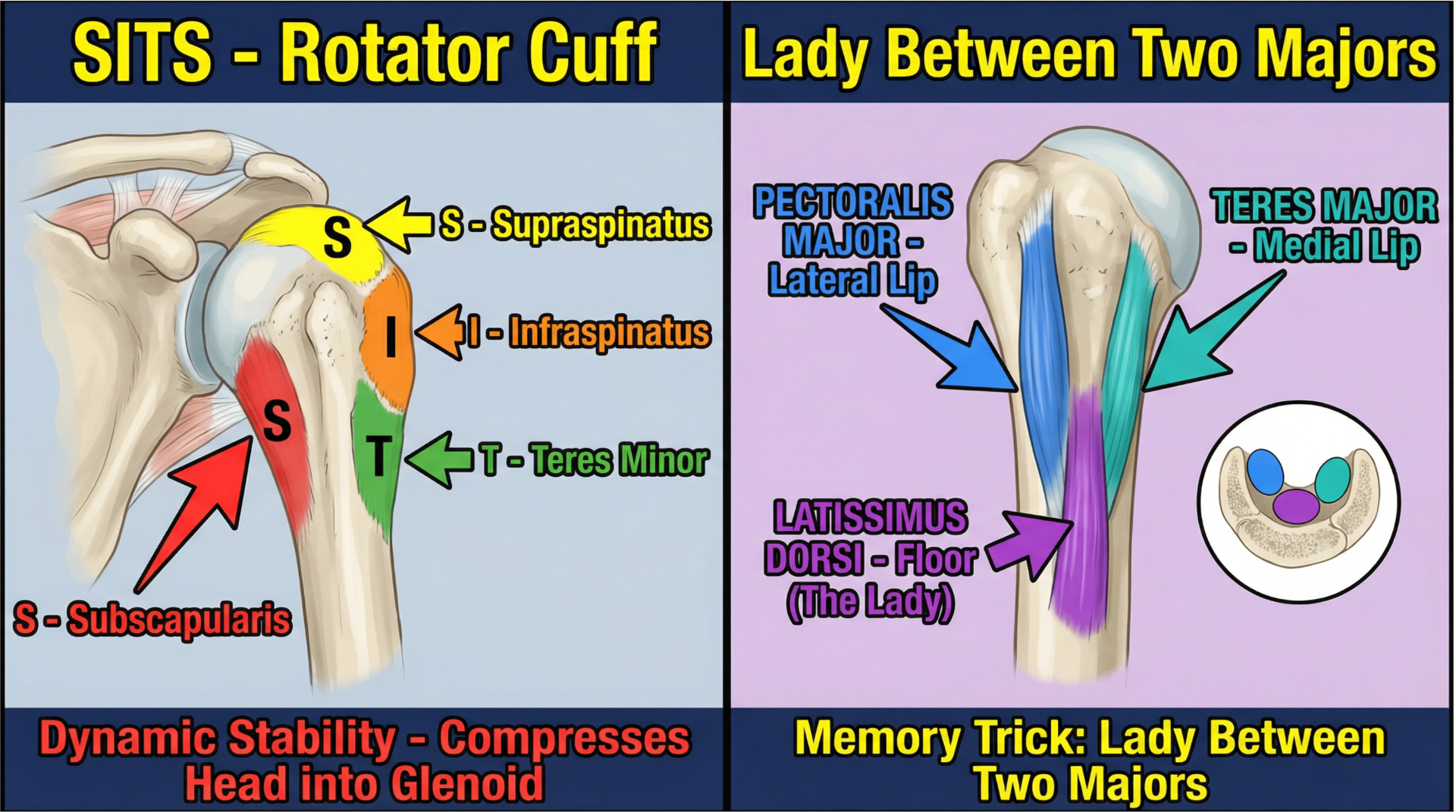
The Rotator Cuff (SITS)
Comprising the SITS muscles, these insert onto the tubercles to provide dynamic stability by compressing the humeral head into the glenoid fossa.
"Lady Between Two Majors"
A mnemonic for the bicipital groove attachments:
5. Clinical Kinesiology: Nerve Relationships
Critical areas where nerves contact the bone — vulnerable during fractures
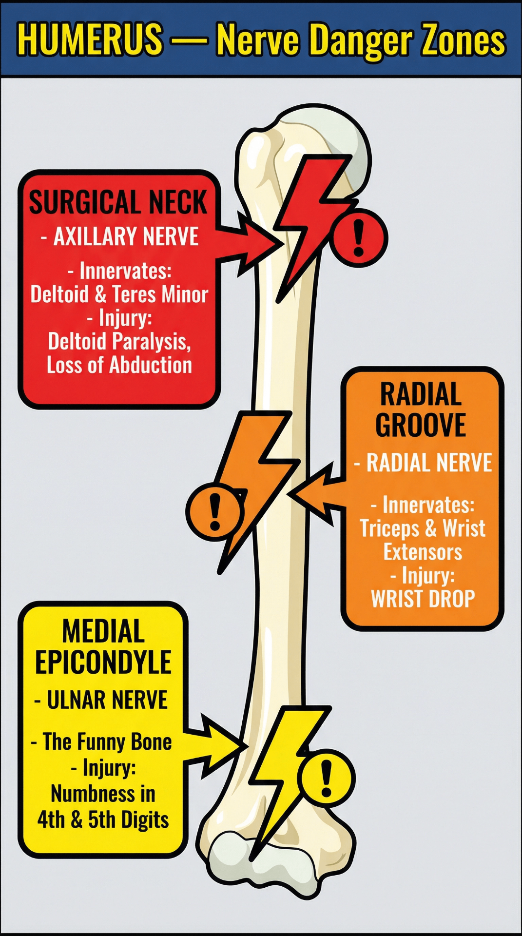
Axillary Nerve
Innervated muscles: Deltoid and Teres Minor
Fracture here can lead to paralysis of the deltoid, resulting in an inability to abduct the arm.
Radial Nerve
Innervated muscles: Triceps brachii and wrist/finger extensors
Fracture of the mid-shaft can cause "wrist drop" due to paralysis of the extensor muscles.
Ulnar Nerve
Innervated muscles: Flexor carpi ulnaris and medial half of flexor digitorum profundus
Injury here can cause numbness and tingling in the 4th and 5th digits (the "funny bone" sensation).
6. Practice Calculation: Elbow Flexion Torque
Consider the Brachialis muscle. If it generates a tension force Fm at an insertion angle θ relative to the ulna, the effective torque τ at the elbow is:
τ = r · Fm · sin(θ)
r — distance from the elbow joint axis to the insertion on the ulnar tuberosity.
θ — changes throughout the range of motion, reaching peak mechanical advantage when the elbow is at approximately 90°.