Radius
An anatomical and biomechanical study guide focusing on the lateral bone of the forearm — the mobile bone that pivots around the ulna to facilitate pronation and supination.
1. Osteology of the Radius
Divided into proximal end, shaft (diaphysis), and distal end
| Region | Landmark |
|---|---|
Proximal | Head |
| Neck | |
| Radial (Bicipital) Tuberosity | |
Shaft | Anterior Border |
| Posterior Border | |
| Interosseous Border | |
Distal | Ulnar Notch |
| Styloid Process | |
| Dorsal (Lister's) Tubercle | |
| Articular Facets |
2 & 3. Muscular Attachments
Muscles originating from and inserting onto the radius
These muscles have their proximal attachment on the radius and typically act on the thumb and fingers.
| Muscle | Origin Site |
|---|---|
| Flexor Pollicis Longus | Anterior surface of the radial shaft and interosseous membrane |
| Flexor Digitorum Superficialis (radial head) | Proximal anterior surface (oblique line) |
| Abductor Pollicis Longus | Posterior surface of the radius (and ulna and interosseous membrane) |
| Extensor Pollicis Brevis | Posterior surface of the distal third of the radius |
These muscles originate from the humerus, ulna, or scapula and insert onto the radius, primarily acting on elbow flexion, pronation, and supination.
| Muscle | Insertion Site |
|---|---|
| Biceps Brachii | Radial (bicipital) tuberosity |
| Supinator | Lateral surface of the proximal third of the radius |
| Pronator Teres | Lateral surface of the mid-shaft |
| Brachioradialis | Lateral surface of the distal end, proximal to the styloid process |
| Pronator Quadratus | Anterior surface of the distal quarter of the radius |
4. Memory Tricks & Key Concept
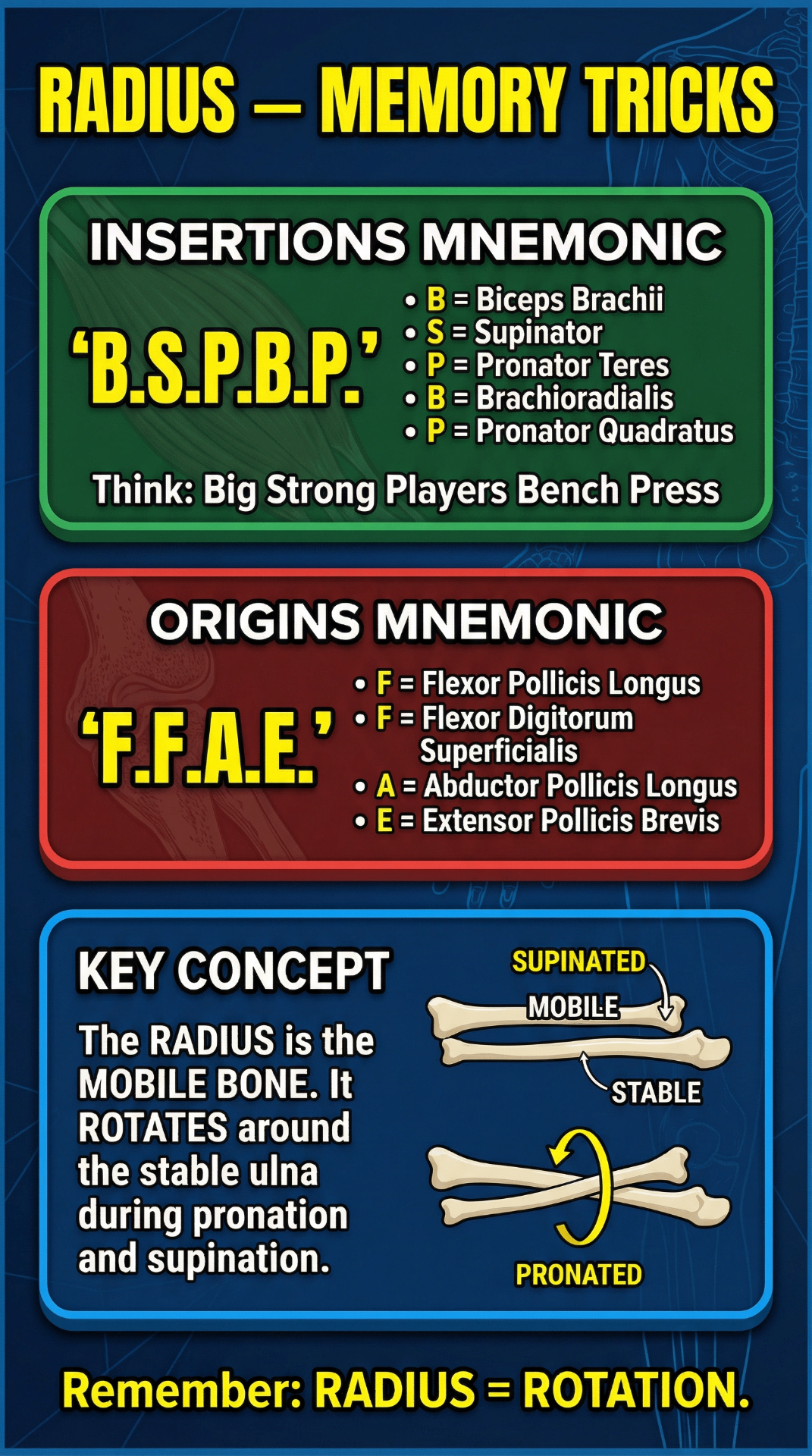
Insertions Mnemonic: B.S.P.B.P.
"Big Strong Players Bench Press"
Remember all 5 muscles inserting onto the radius:
Origins Mnemonic: F.F.A.E.
Only 4 muscles originate from the radius:
Key Concept: Radius = Rotation
The radius is the mobile bone of the forearm. It rotates around the stable ulna during pronation and supination. This is in contrast to the ulna, which remains relatively fixed. Remember: Radius = Rotation.
5. Clinical Notes
Common fracture patterns — FOOSH = Fall On OutStretched Hand
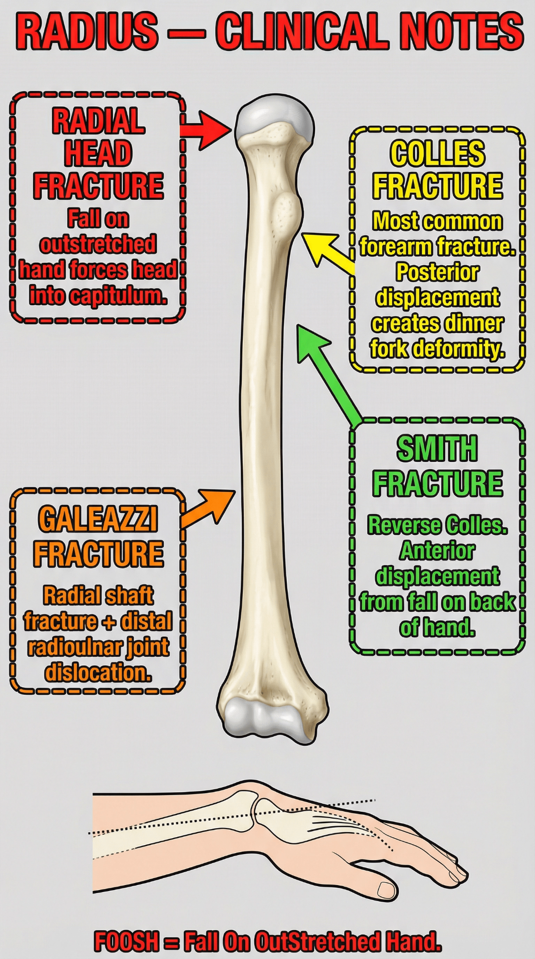
Radial Head Fracture
Fall on outstretched hand (FOOSH) forces the radial head into the capitulum of the humerus.
Often results in pain during forearm rotation. Can be associated with loss of full elbow extension. Common in adults after a FOOSH injury.
Colles' Fracture
The most common forearm fracture. A fracture of the distal radius with posterior displacement of the distal fragment.
Produces the classic 'dinner fork' deformity when viewed from the side. Caused by a FOOSH injury. Most common in elderly individuals with osteoporosis.
Smith Fracture
A 'reverse Colles' fracture. A fracture of the distal radius with anterior (volar) displacement.
Caused by a fall on the back of the hand (a flexed wrist). Less common than Colles' but important to differentiate.
Galeazzi Fracture
A fracture of the radial shaft combined with dislocation of the distal radioulnar joint (DRUJ).
A serious injury that typically requires surgical fixation. The counterpart to the Monteggia fracture of the ulna.