Ulna
An anatomical and biomechanical study guide focusing on the stabilizing bone of the forearm — its bony landmarks, muscular attachments, and clinical significance.
1. Osteology of the Ulna
Divided into proximal epiphysis, diaphysis (shaft), and distal epiphysis
| Region | Landmark |
|---|---|
Proximal | Olecranon Process |
| Trochlear Notch | |
| Coronoid Process | |
| Radial Notch | |
| Ulnar Tuberosity | |
Shaft | Anterior Border |
| Posterior Border | |
| Interosseous Border | |
Distal | Head of Ulna |
| Styloid Process |
2 & 3. Muscular Attachments
Muscles originating from and inserting onto the ulna
These muscles have their proximal attachment on the ulna and typically act on the wrist, hand, and fingers.
| Muscle | Origin Site |
|---|---|
| Flexor Carpi Ulnaris | Olecranon process and posterior border of the ulna |
| Pronator Teres (ulnar head) | Coronoid process |
| Flexor Digitorum Profundus | Anterior and medial surfaces of the ulna |
| Extensor Carpi Ulnaris | Posterior border of the ulna (via a shared aponeurosis) |
| Supinator | Ulnar crest (supinator crest) below the radial notch |
| Abductor Pollicis Longus | Posterior surface of the ulna (and radius and interosseous membrane) |
| Extensor Pollicis Longus | Posterior surface of the middle third of the ulna |
| Extensor Indicis | Posterior surface of the distal third of the ulna |
These muscles originate on the humerus or scapula and insert onto the ulna, primarily acting on the elbow joint.
| Muscle | Insertion Site |
|---|---|
| Triceps Brachii | Olecranon process |
| Anconeus | Lateral aspect of the olecranon and posterior proximal ulna |
| Brachialis | Ulnar tuberosity and coronoid process |
4. Memory Tricks & Key Concept
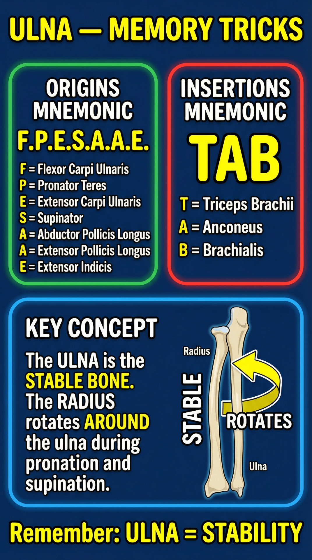
Origins Mnemonic: F.P.E.S.A.A.E.
Remember all 7 muscles originating from the ulna:
Insertions Mnemonic: TAB
Only 3 muscles insert onto the ulna — easy to remember!
Key Concept: Ulna = Stability
The ulna is the stable bone of the forearm. During pronation and supination, the radius rotates around the ulna. The ulna remains relatively fixed, providing a stable axis for the forearm's rotational movements.
5. Clinical Notes
Important clinical correlations — fracture zones and nerve vulnerabilities
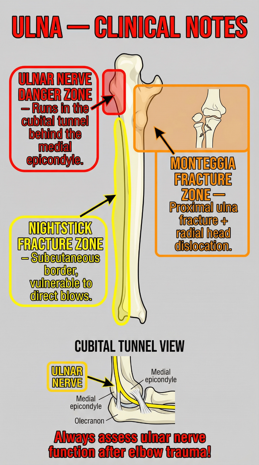
Ulnar Nerve Danger Zone
The ulnar nerve runs in the cubital tunnel behind the medial epicondyle, directly adjacent to the olecranon.
Compression or trauma here causes 'cubital tunnel syndrome,' leading to numbness and tingling in the 4th and 5th fingers and weakness in hand grip.
Nightstick Fracture Zone
The subcutaneous posterior border of the ulnar shaft is vulnerable to direct blows.
A direct blow (e.g., raising the arm in defense) can cause an isolated fracture of the ulnar shaft, known as a 'nightstick fracture.'
Monteggia Fracture
A complex injury involving the proximal ulna and the radioulnar joint.
A fracture of the proximal third of the ulna combined with dislocation of the radial head. This is a serious injury that requires surgical intervention.